601.483.0011 |
601.483.0011 |
 What Is Digital Mammography?
Mammography (also known as a mammogram procedure) is a specific type of imaging that uses a low-dose X-ray system for the examination of breasts. A mammography exam is used as a screening tool to detect early breast cancer in women experiencing no symptoms and to detect and diagnose breast disease in women experiencing symptoms such as a lump, pain or nipple discharge.
Mammography plays an important part in the early detection of breast cancers because it can show changes in the breast up to two years before a patient or physician can feel them. Current guidelines of the U.S. Department of Health and Human Services (HHS), the American Cancer Society (ACS), the American Medical Association (AMA) and the American College of Radiology (ACR) recommend a screening mammography every year for women, beginning as early as age 40. Research has shown that annual mammograms lead to early detection of breast cancers when they are most curable and breast-conservation therapies are available.
The National Cancer Institute (NCI) adds that women who have had breast cancer and those who are at risk due to a genetic history of breast cancer should seek expert medical advice about whether they should begin screening before the age of 40 and about the frequency of screening.
If you are or suspect that you might be pregnant, let your doctor, nurse or technologist know as soon as possible. The American Cancer Society also recommends you:
Same-day results through MyOchsner.
Imaging Center Hours
Monday–Thursday 7:30 a.m.–4:30 p.m.; Friday 7:30 a.m.–11:30 a.m.
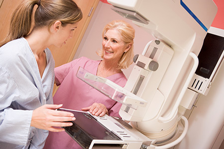
A licensed radiologic technologist will perform your exam. Your breast will be placed on a special platform and compressed with a clear plastic paddle.
Breast compression is necessary to:
The technologist will stand behind a glass shield during the X-ray exposure. You will be asked to change positions slightly between images. The routine views are a top-to-bottom view and an oblique side view. The process will be repeated for the other breast.
The examination process should take about half an hour. When the procedure is completed, you will be asked to wait until the technologist examines the images to determine if more are needed.
You will feel pressure on the breast as it is squeezed by the compression paddle. Some women with sensitive breasts may experience discomfort. If this is the case, schedule the procedure when your breasts are least tender. The technologist will gradually compress your breast. Be sure to inform the technologist if pain occurs as compression is increased. If discomfort is significant, less compression will be used.
Our facility is recognized as an FDA-approved mammography site, and our services are accredited by the American College of Radiology. Your mammogram will be interpreted by board-certified radiologists. Mammogram results will be available the same day through MyOchsner for you and your provider. Also, a letter containing the results of your mammogram will be mailed to your home address.
For more information, please call 601.703.9520.